Neurons, songbirds and candy-coated science: Young minds explore neurobiology
Fourth- and fifth-grade students near USC’s University Park Campus recently had the unusual opportunity to dive into neurobiology through a series of hands-on lessons inspired by research at the USC Dornsife College of Letters, Arts and Sciences.
Professor of Biological Sciences and Psychology Sarah Bottjer, whose research on neural anatomy and vocal learning in songbirds inspired the lessons, led young students at Vermont Avenue Elementary School through a series of activities, aided by staff from the Joint Educational Project based at USC Dornsife.
After a brief presentation of Bottjer’s research, students formed small groups to explore brain anatomy, neuron signaling and how songbirds learn to sing. The workshop combined music, art and science to make complex topics understandable and memorable, enabling the students to spend some time the shoes of neuroscientists.
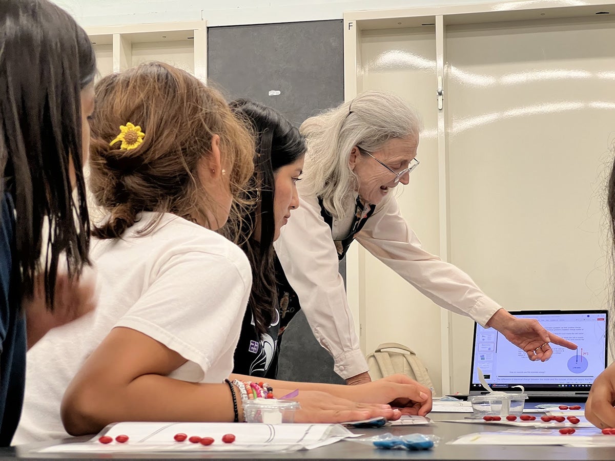
Candy-coated Ions: USC Dornsife neuroscientist Sarah Bottjer taught students about concentration gradients using M&M candies. The colorfully coated chocolates, which represented ions that are key to communication between neurons, enabled the students to see how neurons relay electrical signals to muscles. (Photo: Kathrin Rising.)
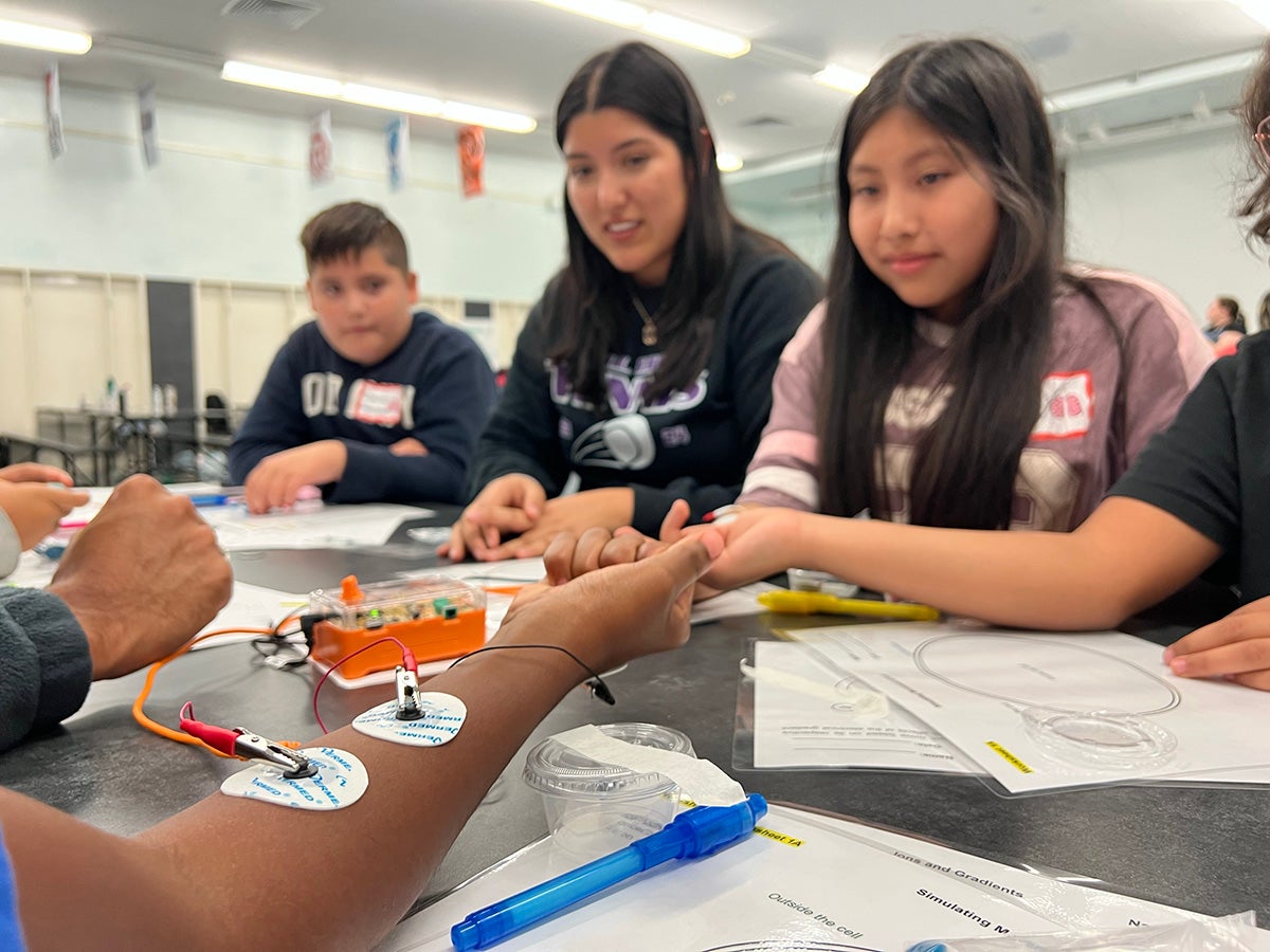
Electrical Connection: Students saw how the brain sends signals to muscles using a device called a Human-to-Human Interface (HHI). The HHI detects brain signals from one student and carries them to another. One student moved her arm with electrodes attached, and the HHI sent a neural signal to electrodes taped to another student’s arm causing it to move. (Photo: Kathrin Rising.)
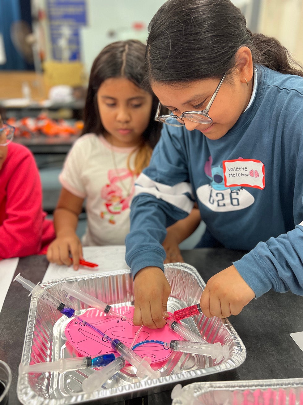
Follow the Glow: Students explored how scientists use fluorescent and protein tracers to map neuron pathways in the brain. Using a laminated paper brain, tubing and syringes filled with food coloring, students simulated the movement of a fluorescent tracer. (Photo: Dieuwertje Kast.)
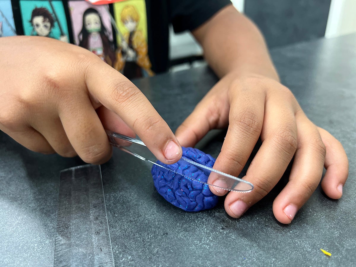
Think Thin: Students simulated a cryostat — an apparatus for taking very thin slices of deeply frozen tissue — by cutting a Play-Doh brain into 1-millimeter slices. (Photo: Dieuwertje Kast.)
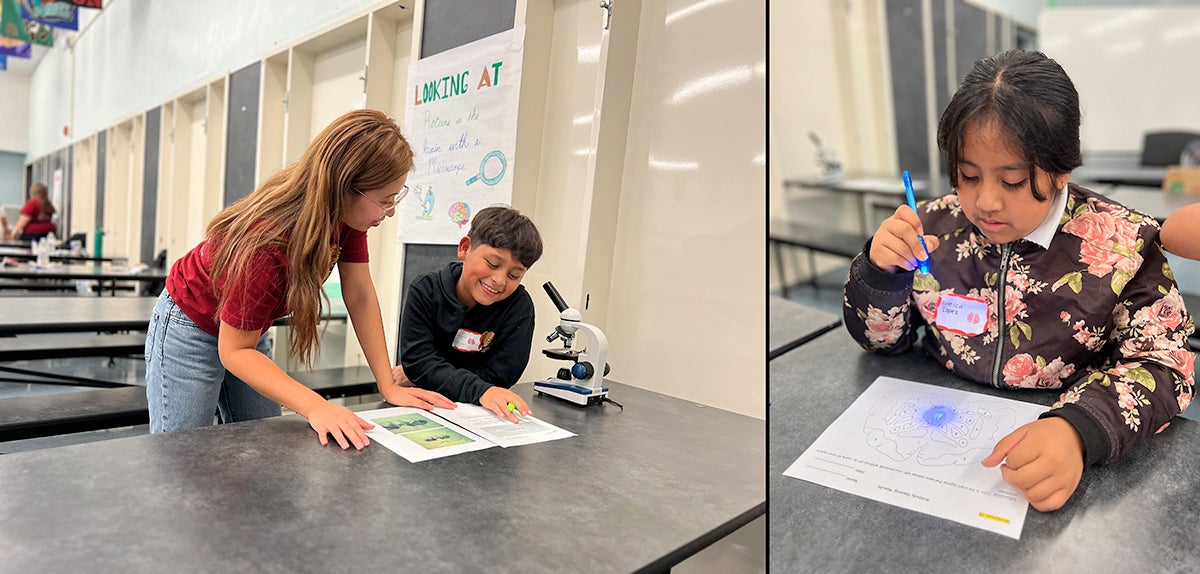
Seeing Is Believing: At left, USC Dornsife biological sciences major Keira Zoleta of JEP’s STEM Education Programs showed a young student a real brain slice using a microscope. At right, a student learned how antibody staining works in the brain by using invisible ink to label different proteins on a worksheet. (Photos: Kathrin Rising and Dieuwertje Kast.)
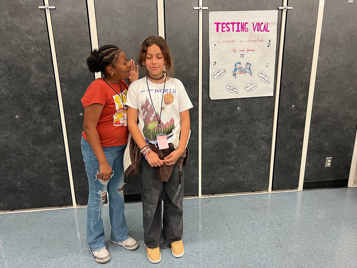
Tuning Into Learning: Using familiar tunes like “Twinkle, Twinkle, Little Star” and “Happy Birthday to You,” students learned how young songbirds raised by different “foster parents” learn the foster parents’ songs. This experiment, a highlight for many of the students, demonstrated that behavior can be learned rather than innate. (Photo: Kathrin Rising.)
