A new microscopy facility at USC opens a world of possibilities for scientists
About 500 years ago, a Dutch maker of spectacles first paired two refractive lenses to magnify objects in his view, revealing a world so unexpected and new it could only be described by the instrument that revealed it. Ever since, scientists have endeavored to peer ever deeper into the microscopic universe, inventing new instruments and techniques to expand their view of this infinitesimal world.
The benefits have been innumerable, particularly in the fields of biology and human health, as microscopic organisms, then individual cells and then cellular components came into focus. Now, even individual molecules can be visualized, thanks to electron microscopes.
Imaging molecules has become increasingly important to a branch of science called structural biology. Knowing the shape of biological molecules such as proteins and DNA, and how their shapes change as they interact with other molecules, is key to understanding how they function within cells. And it can lead researchers to new drug targets.
A cool development in microscopes
One of the most significant advances in structural biology builds on the power of electron microscopy by cooling samples to extremely low temperatures approaching absolute zero, where molecules themselves stop moving altogether. Called cryo-electron microscopy, or cryo-EM, the technique enables scientists to take a snapshot of biological molecules in three dimensions — and it does so far more quickly than traditional methods.
“It really is a game changer in biology,” said Stephen Bradforth, divisional dean for the physical sciences and mathematics at the USC Dornsife College of Letters, Arts and Sciences. “It enables us to very quickly do what used to be impossible.”
And now USC will be part of that game change thanks to a co-location agreement with Amgen, one of the world’s largest biotechnology companies, which has resulted in not one, but two, of the world’s most advanced cryo-EM systems being housed at the university.
Creative solutions to a necessary expense

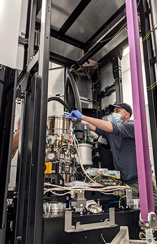
A technician adjusts cryo-EM components during installation.
Cryo-EM has become so important to structural biology research that some funding organizations, when deciding whom to award grants, are beginning to favor institutions that have direct access to the equipment.
“Grant reviewers will say, ‘Well, this proposal doesn’t have an attempt to collaborate with somebody doing structural biology, so therefore I’m not going to fund it,’” explains Bradforth, professor of chemistry at USC Dornsife. “And suddenly everybody who’s doing biology realizes they’ve got to have some way to get access to this.”
But there’s a catch: The technology comes with a significant financial burden. The least expensive systems cost millions of dollars, and that doesn’t factor in the cost of building a facility capable of housing the sensitive instruments. In addition, they require a highly skilled person or team to operate them.
In short, ownership of a cryo-EM system is beyond the financial means of many academic research institutions. And despite the technology’s value in searching for potential new therapeutic drug targets, even biotechnology or pharmaceutical industry giants may have difficulty justifying some of the expense to their shareholders.
But the USC-Amgen co-location agreement solved this problem by combining the company’s capital with the university’s commitment and expertise to run the instruments as well as space in which to locate them.
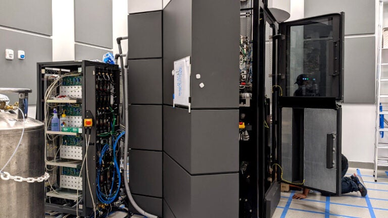
Designed and manufactured by Thermo Fisher Scientific Inc., the Glacios and Krios cryo-EM instruments reside in the USC Michelson Center for Convergent Bioscience, where scientists from both USC and Amgen will share access.
The collaboration is part of a growing trend in research, says Amgen’s Philip Tagari, vice president of research.
“We are seeing the ‘century of biology’ unfolding. Advances are coming at unprecedented speed, as recently demonstrated by the pace of innovation related to COVID-19,” Tagari said. “The biopharmaceutical industry, from ‘virtual’ startups to established major pharmaceutical companies, will need to partner with world-class academic institutions in order to best translate new discoveries into breakthrough therapies for unmet medical needs.”
A font of research opportunities
Scott Fraser is a renowned biological imaging expert who leads USC’s Translational Imaging Center, a core facility at the university focused on creating new biological imaging technologies.
Fraser, Provost Professor of Biological Sciences, Biomedical Engineering, Physiology and Biophysics, Stem Cell Biology and Regenerative Medicine, Pediatrics, Radiology and Ophthalmology at USC, sees the new cryo-EM facility as a central core for problem-solving.
“Somebody’s got a problem … and they have no idea how to solve it, and they come to a facility like this with people that know about the field, and they go from a problem to a solution in maybe one-tenth the amount of time it would have taken on their own,” he said.
The benefits are both intellectual and financial, he says, noting that in a recent audit of the Translational Imaging Center, reviewers found that “for every dollar spent on that core, there were between $4 and $5 of new research grants generated.”
Cornelius Gati leads the new cryo-EM core facility at USC. He joined the university in December 2020 as assistant professor of biological sciences after honing his cryo-EM skills at MRC Laboratory of Molecular Biology in the United Kingdom and Stanford University.
Gati is excited by the potential for accelerating USC’s research efforts with the new, in-house core facility, as well.
“There are lots of structural biologists at USC who have been collaborating with people outside of the university because there were no instruments here yet,” he said. Now, in much the same way those first microscopes transformed science, this new, on-campus facility opens a universe of possibilities for USC’s research efforts.