Layer By Layer
Part meticulous study, part eloquent homage, Leonardo da Vinci’s Vitruvian Man is considered not only a seminal work of art, but the very embodiment of the boundary-breaking, outward-looking curiosity of the Renaissance period.
Based on the work of a first-century Roman architect, Vitruvius, the intricate sketch of a man — arms outstretched, torso floating within a circle balanced neatly within a square — is a poetic rendering of symmetry and proportion. However, what the image and accompanying text most vitally illustrate is an intrinsic bridge between the arts and sciences. It underscores that humans are microcosms of the universe itself. “Man,” da Vinci wrote, “is the model of the world.”
Five hundred years later this rendering continues to loop through the minds of a group of leading biomedical researchers at USC Dornsife — both as a challenge and an inspiration.
“One of the biggest scientific accomplishments in history was da Vinci’s Vitruvian Man,” began Raymond Stevens, Provost Professor of Biological Sciences and Chemistry. “Although da Vinci originally created the painting for the sake of art, he was one of the first to create a map of the human body and started a path toward deciphering everything we are made up of.”
“The connection between the ‘everything’ Ray just referred to — biology, chemistry, engineering, medicine — has long since frayed,” added Peter Kuhn, Dean’s Professor of Biological Sciences. “This approach has been replaced by modes of inquiry characterized by highly individualized research and specializations. Scholars often work in isolation.”
Since da Vinci’s time, the sciences in particular have diverged into myriad subdisciplines.
“While this splintered approach has led to significant advances in our fundamental understanding of the world,” explained Scott Fraser, Provost Professor of Biological Sciences, “it has also resulted in silos of expertise that run deep and often aren’t adequate in addressing the complex challenges we now face, especially those in human health.”
USC Dornsife has established The Bridge@USC as an antidote to this silo culture.
Stevens, Kuhn and Fraser are among a founding group of top USC scientists who have joined forces to set a new paradigm for how 21st-century research is conducted and applied.
In launching The Bridge@USC, they are uniting outstanding minds in chemistry, biology, medicine, mathematics, physics, engineering and nanoscience — as well as experts in such areas as animation and cinematography — to build the first atomic-resolution model of man.
The creation of this dynamic, virtual model, USC Dornsife Dean Steve Kay asserted, will accelerate the development and implementation of innovative therapies and cures for a host of intractable diseases and conditions such as cancer, Alzheimer’s, Parkinson’s and diabetes.
“We are forging partnerships with schools across the university to create a bridge among different types of researchers: engineers, scientists, artists, medical doctors,” Kay said. “Everybody wants to work together, but it’s often difficult for a variety of reasons ranging from funding to different scientific languages and data types. So we are helping to provide the unifying framework that makes this possible.”
The university’s support of such entrepreneurial endeavors is exactly what attracted this cluster of pioneering scientists to USC.
Stevens, who earned his Ph.D. in chemistry from USC Dornsife in 1988, returned in Fall 2014 and serves as founding director of The Bridge@USC. He is joined by associate directors Kuhn and Vadim Cherezov, both of whom were his former colleagues at The Scripps Research Institute, along with Vsevolod “Seva” Katritch, assistant professor of biological sciences, and former Cold Spring Harbor Laboratory collaborator James Hicks, professor (research) of biological sciences. This August, Valery Fokin, who was also previously at Scripps and earned his Ph.D. in chemistry from USC Dornsife in 1998, arrives as professor of chemistry.
“A group of like-minded faculty, supportive and dynamic administrators, and visionary and generous supporters — it is a sine qua non for that to happen,” Fokin said. “A combination of all three is a rarity, and USC is at this unique point now.”
Together their laboratories bring a cohort of approximately 70 researchers to the university.
In addition, Kay and Fraser have joined the initial Bridge@USC founding team, which will grow to include eight additional faculty hires over the next three years. They will also continue to forge partnerships with collaborators from across USC Dornsife, the USC Viterbi School of Engineering, Keck School of Medicine of USC and USC School of Cinematic Arts.
Spanning Disciplines
Centuries since da Vinci’s detailed rendering of the human form, we remain fascinated by the body — how it works and how it fails. And while we have categorized most of the elements of man at the genetic, molecular and cellular levels, Kuhn pointed out, we have yet to integrate the different scales of data together.
“Until those gaps are spanned,” he said, “the most effective and efficient ways to develop new drugs and understand diseases will continue to confound and outpace us.”
This work, however, isn’t occurring in a vacuum. The European Commission has launched the Human Brain Project, which aims to deliver a “scaffold” model of the human brain in the next decade, and President Barack Obama has created the BRAIN (Brain Research through Advancing Innovative Neurotechnologies) Initiative. Google recently announced that it has embarked on a quest to create a more complete picture of the human brain, hoping to pinpoint how diseases might be prevented rather than merely treated. While theirs is a “top down” approach, The Bridge@USC aims to do the opposite, working up from the molecule to the cell to the entire human body.
“I think our uniqueness is that we combine structure on the human, cellular and molecular scales. Both static and dynamic structure tools are available at all levels and the time is now right to pull this together,” said Stevens, who holds joint appointments at USC Viterbi and the Keck School.

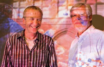
Peter Kuhn (left) and James Hicks (right) have devised a way to test for cancer cells in the blood through what they have dubbed “a liquid biopsy.” In addition to being less costly and uncomfortable for patients, the method may also be more sensitive and effective at identifying circulating tumor cells than any of their existing competitors. Photo by Ryan Young.
“What is incredibly exciting is the opportunity to work with the digital arts faculty in the USC School of Cinematic Arts who are ranked No. 1 in the world, and the USC Institute of Creative Technologies. Furthermore, we are excited to work with and enable our colleagues at USC Viterbi and the Keck School with the breakthrough information that comes out of this endeavor.”
The team’s first step, though, has been to determine how their areas of expertise best correspond to the targeted layers of the bottom-up study, which will ultimately allow doctors to better detect and treat human disease.
Construction of The Bridge@USC’s virtual model of the human body needs to be approached from three different levels simultaneously — molecules, cells and whole body — while connecting the different scales together, Stevens explained.
Level 1: Molecules
Molecules are formed when two or more atoms join together chemically. Stevens and Cherezov, professor of chemistry, image molecules, particularly the proteins in the lipid membrane involved in cellular communication, to see how individual proteins bind with signaling molecules or drug candidates. Katritch’s expertise is in developing and applying computational tools to study key biological phenomena — including virtual drug screening and understanding the molecular basis of drug action. Katritch then uses computer
modeling to infuse potential drug treatments into those protein-binding sites that Stevens and Cherezov have observed.
To next synthesize new compounds that most effectively target specific diseases, all three benefit from Fokin’s click chemistry methodology that allows them to develop new chemical probes — substances that alter specific protein function — and better understand receptors in the human body. This chemistry work is complemented beautifully by that of USC Dornsife chemists Charles McKenna, Surya Prakash and Nicos Petasis.
Stevens, Cherezov and their research teams have already unlocked the biomedical potential of several G protein-coupled receptors (GPCRs) by determining their structure. GPCRs serve as the cell’s gatekeepers and messengers, receiving and sending information in the form of light energy, peptides, lipids, sugars, and proteins. Their signals mediate practically every essential physiological process, from immune system function to taste and smell to cognition to heartbeat.
With nearly 1,000 members, GPCRs constitute the largest protein family in the human genome — and a key avenue to medical progress. These receptors are responsible for 80 percent of cell membrane signaling; some 40 percent of all pharmaceuticals act by binding to GPCRs.
The techniques developed by Cherezov have enhanced the biophysical characterization and crystallization of membrane proteins fostering a revolution in structural studies of GPCRs, whose malfunctions often result in a range of diseases and conditions. He likens his approach to that of Eadweard Muybridge, whose experiments with motion photography in the late 1880s proved that contrary to popular belief all four of a horse’s hooves do leave the ground at once when it gallops. Cherezov has developed numerous novel instruments and technologies that should eventually allow scientists to see molecules in motion and observe changes in proteins as they occur.
Cherezov sees the magnitude of The Bridge@USC’s goal to create an atomic-resolution model of man as more complex compared to the ambitious Human Genome Project in the 1990s.
“With the scientific field moving so fast, although it sounds absurdly ambitious, it is now feasible for us, with all of the tools and data available, to visualize the structure and dynamics of individual molecules, to build blocks of the cell, and then to start assembling them together in space and time,” Cherezov said.
Utilizing structural bioinformatics and integrative molecular modeling approaches to decipher the intricate mechanisms of GPCR signaling, Katritch identifies new venues to precisely modulate GPCRs by ions and small molecules, leading to better treatment.
“What we are trying to do,” Katritch said, “is apply a systemic approach to study the whole GPCR family, to compare them and to figure out how they work based on combining and bridging structural, biochemical and biophysical knowledge. There are at least 826 receptors, making up a significant chunk of the human genome, and each has its own character and a distinct role in human biology and disease.”
The therapeutic potential for patients with immune and metabolic diseases is vast. Stevens expects the immediate impact of this work to be in diabetes, heart disease, cancer, embryonic development, and neurodegenerative diseases such as Alzheimer’s and Parkinson’s.
Level 2: Cells
Molecules — billions of them — then make up cells, the essential building blocks of all living organisms. Each cell acts as a house that organizes the molecules’ functions and determines how these will communicate with other cells to create tissues, organs and whole organisms. When something goes awry on the molecular level, this affects the cells, which can impact tissues, organs and the whole body.
The realm of the cellular is Fraser’s expertise. He specializes in imaging the fine details from the cellular level all the way to the organs.
Fraser collaborates with biologists, engineers, chemists and physicians to build new technologies for imaging biological structures and function. These state-of-the-art devices allow researchers to explore, in real time, the inner workings of such complex events as embryonic development and disease progression. By better observing the basic behaviors of cells, Fraser strives to improve regenerative, preventive and personalized medicine.
For example, he constructs microscopes that allow scientists to watch as cells interact with one another to form the heart muscle and valves. Understanding this process — how cells give off signals, respond and collaborate to build an embryonic heart — may offer keys to rebuilding heart valves in vitro.
“With USC’s recruitment of Arthur Toga and Paul Thompson from UCLA to image the brain, Andrew McMahon from Harvard University to focus on stem cells, and Stevens, Fokin, Kuhn, Cherezov, and Katritch from Scripps, I am in a perfect situation to realize a scientific dream of connecting molecules to man at the atomic level,” said Fraser, who holds joint appointments at USC Viterbi and the Keck School.

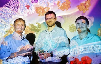
Through their advances in human cell signaling, Raymond Stevens (left), Vadim Cherezov (center) and Vsevolod “Seva” Katritch (right) are helping scientists to design pharmaceuticals that more effectively target diabetes, cancer, and mental disorders. Photo by Ryan Young.
Level 3: Body
Made up of 78 organs and networks including the brain, heart, lungs and gastrointestinal tract, the body — the intricate physical structure of the human form — is where Kay, Kuhn and Hicks are focused.
There has been a wellspring of research about how circadian rhythms affect health and overall well-being, but Kay’s research homes in on how the body’s timing of the day/night cycle can influence the onset of diabetes and obesity. His laboratory deploys advanced imaging techniques, uses computational approaches to understanding the dynamics of physiological networks, and takes full advantage of an array of next-generation sequencing and chemical biology tools to illuminate the complexities of metabolic regulation.
He found that a key protein, cryptochrome — which regulates the biological clocks of plants, insects and mammals — also regulates glucose production in the liver. Kay and his collaborators observed that altering the levels of this protein could improve the health of diabetic mice. Like mice and other animals, humans have evolved complex biochemical mechanisms to keep a steady supply of glucose flowing to the brain at night, when we’re not eating or active.
More recently, Kay and his team used high throughput screening to discover a novel small molecule, KL001, which controls the intricate molecular cogs or timekeeping mechanisms of cryptochrome in a way that can repress the production of glucose. This finding opens potentially groundbreaking avenues for the development of drugs to treat diabetes and other metabolic disorders. The serendipitous discovery occurred during a parallel effort in Kay’s laboratory to identify molecules that regulate the periodicity of the biological clock in predictable ways.
“Our next aim is to understand how KL001, and similar molecules that affect cryptochrome, function in whole animals,” said Kay, who holds joint appointments at USC Dornsife and the Keck School. “We are going to investigate how such compounds affect other processes besides the liver as we believe our work holds promise not only for diabetes, but also for diseases such as asthma and some cancers.”
By examining the circulatory system and detecting how molecules traverse the body, both Kuhn and Hicks are zeroing in on a better understanding of how unwanted molecules or single cells might cause diseases such as cancer, particularly with the power of single cell genomics. And they point to the distinct advantage they have at USC because of the strength of its computational genomics program — led by University Professor Michael Waterman and Andrew Smith, associate professor of biological sciences.
“The beauty of the bloodstream is that it’s a super highway that connects the entire body,” said Kuhn, who holds joint appointments at USC Viterbi and the Keck School. “A cancer cell that breaks away from the primary tumor gets exposed to the whole body through the circulatory system in just one minute — the time it takes for blood to circulate.” Kuhn decided to exploit that super highway, believing that analysis of cancer cells in the blood can be a complement to traditional imaging techniques that provide information about the tissue parts of tumors.
How cancerous cells gain the ability to exit tumors and populate distant organs is a fascinating yet poorly understood biological question of immense clinical importance. Kuhn has set out to find that “needle in a haystack” by working with oncologists, a mathematics modeling group, and a single-cell genomics group led by Hicks.
Their subsequent method for detecting cancer cells with just a blood sample has yielded a minimally invasive, inexpensive test that differentiates circulating tumor cells (CTCs) — which break away from the primary tumor to metastasize to other parts of the body — from ordinary blood cells using a digital microscope and image-processing algorithm. This advance is expected to achieve results comparable to surgical biopsies without having to submit patients to the operating table. It also significantly enhances doctors’ abilities to detect, monitor and predict cancer progression at an earlier, more treatable stage.
Kuhn and Hicks already are using technology developed in their labs to build a complete high-content model of cells, cellular content and non-cellular content using genomics, proteomics and large-scale computing with their colleagues Paul Newton at USC Viterbi and Jorge Nieva at the Keck School. This breakthrough enables them to identify clinically useful biomarkers and to advance the use of a noninvasive fluid biopsy, which can help inform a doctor and patient’s treatment decisions.
A Larger Purpose
While Stevens and his colleagues are carefully assembling The Bridge@USC’s team, just as crucial is the design of their workspace.
The Bridge@USC will be located in the new 190,000- square-foot USC Michelson Center for Convergent Bioscience, which will support up to 24 principal investigators with laboratories employing hundreds of researchers and students. A bold new collaboration between USC Dornsife and USC Viterbi, the center will feature state-of-the-art, flexible labs that accommodate the spectrum of scientific activities within the broad area of molecular science and engineering and can be reconfigured as needed to adapt to future discoveries. The floorplans are designed with flow and synergy in mind: There are meeting spaces, common areas, even a literal bridge connecting wings.
Mirroring the body’s busy interconnected network, The Bridge@USC will be the kinetic hub that encourages unexpected opportunities for the researchers within the Michelson Center and across USC’s University Park and Health Sciences campuses to cross, even collide. The team believes this possibility for serendipity — stumbling upon unforeseen breakthroughs by accident — is at the heart of scientific inquiry.
Consequently, The Bridge@USC’s goals include fostering unexpected yet promising partnerships through its catalyst program, start-up incubator, venture fund, and academe and industry collaborations.
Stevens, Kay and Fraser have studied the handful of great research institutes and have designed The Bridge@USC to build upon those previous successes while avoiding their pitfalls.
“Ensuring freedom to think out of the box, funding high-risk research, and lowering barriers to catalyze collaborations and creativity are critical,” Kay said.

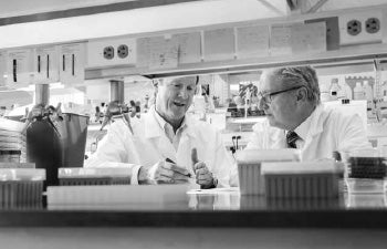
As director of the Translational Imaging Center at USC, Scott Fraser (left) helps fellow faculty members such as Dean Steve Kay (right) accelerate their research by providing access to technologies for the intravital imaging of cells and cellular processes. Photo by Max S. Gerber.
The Catalyst Program will provide seed funding enabling campus teams to collaborate and pursue high-reward, breakthrough research not yet viable to compete for external and government support; a start-up incubator will be developed concurrently with biotechnology and pharmaceutical industry partners; a venture fund will generate new intellectual property and technology transfers; and finally an academe/industry alliance will combine expertise across disciplines to increase and translate the resulting knowledge of the human body.
Led by Stevens, USC has already formed an academe/industry open-source consortium that is generating high-resolution images of at least 200 of the most important GPCRs and investigating the pharmacology of drug interactions. The consortium is creating yet another bridge, this time between USC and the pharmaceutical industry to understand the complete human body.
In addition, The Bridge@USC is committed to providing opportunities for students at the high school, undergraduate, graduate and postdoctoral levels through internships and one-on-one mentoring, and by creating a space where they see themselves not just as scientists, engineers or artists, but as in-the-moment problem solvers. In June, the first Bridge Undergraduate Student program will launch with college and high school students working in The Bridge@USC faculty’s laboratories.
“The sum is bigger than its parts,” Stevens said. “Collaboration and communication — that’s something that has to be a fundamental core value of the institute. That lone-ranger approach to cracking age-old problems is an outdated way of doing science. This is something at universities that has to change because community is key.”
And by community, Stevens means Los Angeles.
While it is too early to predict scientific outcomes, that hasn’t hindered Stevens and his colleagues from thinking broadly, envisioning a larger purpose. The Bridge@USC is only the start — the beginning of a larger plan to build greater Los Angeles into a biotech leader. They have already begun looking beyond the borders of the campus, at the region as whole.
“In terms of biotech and L.A.: Amgen is here and there have been a few recent successes in the L.A. area, but we can do a lot better,” Stevens said. “L.A. is such an ideal place for biotech with large and diverse patient populations, integrated hospital networks and several leading universities. We want to build a stronger biotech ecosystem here to help translate discoveries. The opportunity is just too big from multiple perspectives.”
In other words, The Bridge@USC is not just building an atomic-resolution model of man, but a conversation.
“We’ve designed this to be a nexus, where people throughout the USC campuses can generate a lot of data and can help one another understand their meaning,” Fraser said. “We want to do this in a collaborative, complementary way. A project of this scale requires the cooperation of many different types of scientists, engineers, artists, industries, and governmental agencies. And it’s in such partnerships that the most fruitful advances can occur. That’s critical. It’s all about bridges. It’s coming to the understanding that we’re not going to figure it out by ourselves.”
“The USC Convergent Bioscience Initiative is the modality,” Kay added. “The Michelson Center is the bricks and mortar. And it is the people brought together by The Bridge@USC who will implement the dream — edging us nearer to closing the gaps in knowledge that will help us to better understand the human body. This unique effort includes not only Bridge@USC and affiliated faculty, but our alumni and donors, whose visionary investment will fuel our progress.”
The confluence of expertise, support and location positions USC to be a leader in a modern-day renaissance of scientific inquiry and application — ushering in a new era for L.A.
“Los Angeles should become to medical research what Silicon Valley is to information and technology,” philanthropist and retired orthopedic spinal surgeon Gary K. Michelson, remarked at the center’s groundbreaking last Fall. “We owe it to the world; we owe it to Los Angeles. We need to invest in this.”
Writer Susan L. Wampler contributed to this report.