Mind-Bending Microscope
Thanks to an interdisciplinary, cross-campus effort, USC Dornsife is now home to one of the most advanced microscopes in the world — a device that pushes beyond what was once believed to be the boundary of visible light imaging.
The new GE DeltaVision OMX Blaze can generate 3D images of objects at the nanometer scale, showing crisp details about the structures of cells. Only 58 of these super-resolution microscopes exist in the world, and USC’s is the only one in Los Angeles.
USC’s purchase of the OMX was spearheaded by faculty members Susan Forsburg of USC Dornsife; Scott Fraser, who has joint appointments at USC Dornsife and USC Viterbi School of Engineering; and Noah Malmstadt of USC Viterbi. Support was provided by the Provost Office’s Core Instrumentation Fund, USC Dornsife and USC Viterbi.
“This microscope lends support to USC’s growth in convergent bioscience,” said Randolph Hall, vice president of research at USC. “Our technology will enable life science discoveries, and will lead to new medical devices, materials and therapies designed with incredible precision.”
The new microscope’s future home will be the newly announced USC Michelson Center for Convergent Bioscience, housed at USC Dornsife. It was funded in part by a $50 million gift from philanthropist and retired orthopaedic spinal surgeon Gary K. Michelson and his wife, Alya Michelson.
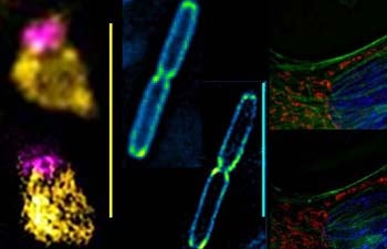
A fission yeast (Schizosaccharomyces pombe) nucleus displaying a pink nucleolar compartment and yellow-histone wrapped DNA; living Shewanella bacteria with peri-cytoplasmic GFP; and mammalian cells labeled with blue nucleus, red mitochondria and green actin cytoskeleton. Top images are conventional epi-fluorescent images, bottom images are the same features in 3D-SIM super resolution. Scale bars in all panels are 5 micrometers. Image courtesy of Marc Green (Susan Forsburg lab).
For now, the OMX resides at the USC Center for Electron Microscopy and Microanalysis (CEMMA) near Zumburge Hall on the University Park campus. It is a shared core instrument. Marc Green, research lab technician at CEMMA, offers training sessions for new users, who can utilize the facility for a modest recharge fee. The cost varies depending on the level of support required, and whether the user is associated with USC.
“We’re keeping the recharge rate low to make this facility as budget-friendly to researchers as possible,” Forsburg said.
The microscope can generate crisp images of objects that are just a few nanometers across — the size of organelles within a cell. Though its ability to take images of living cells with little damage gives it obvious value for biologists, engineers and physical scientists will also find the microscope useful.
“The ability to see dynamic, high resolution detail in samples is invaluable to those of us who are working to develop bio-inspired and smart materials,” Malmstadt said.
Like other such devices, the OMX uses lasers to excite special fluorescent molecules that scientists embed within the object that they wish to image, creating a point of light that can be detected by the microscope.
But no matter how precisely you create a point of light, the fact that it has to pass through a lens — the lens of your eye, the lens of a microscope — means that it will suffer from some sort of diffraction, creating a blurriness around the point’s edges.
That’s where the OMX is different. It gets around the diffraction limit by collecting additional images. These contain interference patterns generated from illuminating the sample through multiple rotating grid patterns (like the Moiré patterns). By analyzing these multiple images with high-powered computer processors, a combined image is generated that pins down, in three-dimensional space, exactly where that point of light is. The resulting images — made up of countless, precisely located points of light — are far sharper than the typical confocal or wide-field deconvolution microscopes in use in USC labs.
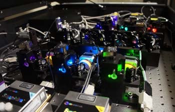
Under the hood of the OMX are six different colored lasers. Photo by Robert Perkins.
With six lasers, scientists can also use differently colored fluorescent probes for different parts of a an object that they want to image — creating a composite image that can be stripped down to individual colors simply by adjusting filters.
And while the laws of physics dictate that an electron microscope, which creates images using electron beams rather than photon beams, will always be able to create images of smaller objects than light microscopes, the OMX has the advantage of being able to image living cells.
“With an electron microscope, you have to use a harsh, chemical fixation process to prepare your samples, which kills the cells,” Forsburg said. “This is suitable for analysis of some cellular structures, but inevitable disrupts or damages others.”
The OMX can be used with live cells, and does not require any special sample preparation process, she said.
“This is a plug-and-play system. You don’t have to specially prepare samples to use in this device — all of your old samples from other light microscope experiments will work just fine,” Forsburg said. Adding to its value, the versatile OMX can also be used in a “standard” mode as a widefield deconvolution microscope, without the super-resolution component.
One of the early adopters of the new microscope is Moh El-Naggar, assistant professor of physics at USC Dornsife, who was recently named as a recipient of the 2012 Presidential Early Career Award for Scientists and Engineers. His research explores structures known as “bacterial nanowires” – nanoscale filaments sent out by bacteria to transport electrons to external surfaces.
“So far we have been limited to either high resolution electron microscopy techniques with non-living bacteria, or low resolution live cell fluorescent techniques,” El-Naggar said. “We no longer have to choose between in vivo and nanoscale resolution. The OMX is the perfect balance.”
By becoming the home to cutting-edge technology, USC will have the opportunity to influence future iterations of that technology.
“The ‘X’ in ‘OMX’ stands for ‘eXperimental,’ which fits perfectly with our attitude of not only being an end-user, but contributing unique applications and even developing new imaging schemes based on this technology,” El-Naggar said.
Researchers interested in using the new OMX can schedule a training session with Marc Green at marcgree@usc.edu.