USC team shows how memories are stored in the brain, with potential impact on conditions like PTSD
Key takeaways:
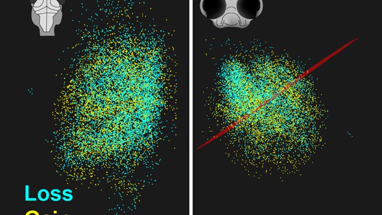
- Contrary to expectations, the study in larval zebrafish shows synapses in one part of the brain are eliminated and new ones are created in a different region when memories are formed.
- These major structural changes could account for memory formation.
- Results may also help explain why unpleasant associative memories, such as those associated with PTSD, are so robust.
- Findings were made possible by a new type of cell labeling and a custom-made microscope designed at USC.
What physical changes occur in the brain when a memory is made?
A team of USC researchers has, for the first time, answered this question by inducing a memory in a larval zebrafish and then mapping changes in their transparent heads with brain cells lit up like Times Square on New Year’s Eve.
After six years of research, they discovered that learning causes brain synapses, the connections between neurons, to proliferate in some areas and disappear in others rather than merely changing their strength, as commonly thought. These changes in synapses may help explain how memories are formed and why certain kinds of memories are stronger than others.
The study was published in the Proceedings of the National Academy of Sciences and was led by a team of faculty from the USC Dornsife College of Letters, Arts and Sciences and the USC Viterbi School of Engineering.
New method and tools
The study was made possible thanks to a new type of cell labeling and a custom-made microscope invented at USC. The researchers also developed a cutting-edge way to track and archive the data collected to make their findings as accessible and reproducible as possible.
The researchers were able to determine for the first time the strength and location of synapses before and after learning occurred in the brain of a living zebrafish, an animal commonly used to study brain function. By taking the unprecedented step of keeping the fish alive, they were able to compare synapses in the same brain over time, a breakthrough in the neuroscience field.
In order to create memories to measure, the research team developed new methods to induce a larval zebrafish to learn. They did this by training the 12-day-old fish to associate a light turning on with being heated on the head with an infrared laser, an action they sought to avoid by swimming away. Fish that learned to associate the light with the impending laser would flick their tails, indicating that they had learned. Five hours of training later, the team was able to observe and capture significant changes in these zebrafish brains.
Surprising results
The main takeaway: Rather than the memory causing the strength of existing synapses to change, the synapses in one part of the brain were destroyed and completely new synapses were created in a different region of the brain.
“For the last 40 years, the common wisdom was that you learn by changing the strength of the synapses, but that’s not what we found in this case,” said Carl Kesselman, a computer scientist at USC Viterbi.
The results suggest that changes in the number of synapses encode memories in the experiment. This also may help to explain why negative associative memories, such as those associated with PTSD, are so robust.
In addition to the findings, USC Dornsife biologist Donald Arnold led a team that created new methods for altering the DNA of the fish so the strength and location of a synapse would be marked with a fluorescent protein that glows when scanned by a laser.
“Our probes can label synapses in a living brain without altering their structure or function, which was not possible with previous tools,” said Arnold.
This made it possible for the specialized microscope developed at USC to scan the brain and image where the synapses were located — without bumping off the fish.
“Sometimes, you try to get such a spectacular image that you kill what you are looking at. For this experiment, we had to find the right balance between getting an image that was good enough to get answers, but not so spectacular that we would kill the fish with photons,” said bioengineer Scott Fraser, who holds appointments at USC Dornsife and USC Viterbi and who led the development of the microscope.
The microscope enabled them to observe changes in living animals and get before and after pictures of the changes on the same specimen. Previously, because experiments were conducted on deceased specimens, they could only compare two different brains, one conditioned, one not.
“This is stealth imaging; we sneak in without being noticed,” Fraser said.
A group led by Kesselman developed innovative new algorithms to process and analyze hundreds of images and experiments.
The study was also unusual for its focus on making the results as transparent and reproducible as possible: All the data associated with the paper is searchable and available at the publicly available Mapping the Dynamic Synaptome website.
“The USC team has set a new bar for data access in that every piece of data generated during the six-year investigation was captured and organized for this research,” said Kesselman, who engineered this new approach.
Added Fraser: “I truly believe that this is the future of transparency in research, a new era, and USC is ahead of the curve.”
Researcher titles:
Don Arnold is professor of biological sciences and biomedical engineering at USC Dornsife and USC Viterbi.
Carl Kesselman is director of the Informatics Division at the USC Information Sciences Institute and professor with the Daniel J. Epstein Department of Industrial and Systems Engineering at USC Viterbi.
Scott Fraser is Provost Professor of Biological Science and Biomedical Engineering at USC Dornsife and USC Viterbi.
About the study
The ongoing study represents the latest cross-disciplinary collaboration at USC with researchers at the USC Michelson Center for Convergent Bioscience, USC Dornsife, USC Viterbi and the Keck School of Medicine of USC. Launched in 2016, the center has brought together a diverse network of premier scientists and engineers under one roof, thanks to a generous $50 million gift from orthopedic spinal surgeon, inventor and philanthropist Gary K. Michelson, and his wife, Alya Michelson.