Researcher hopes to use fluorescence to target tumors
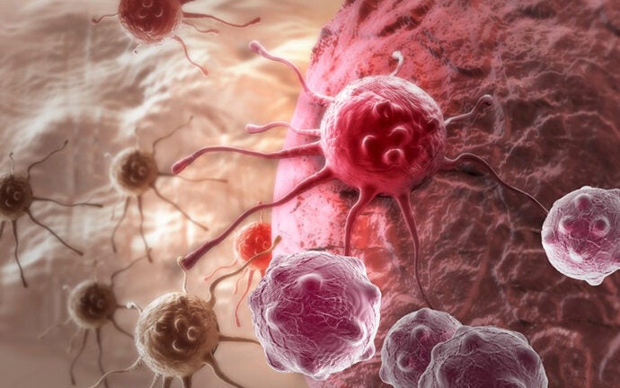
Cancer usually manifests itself in tumors that proliferate quickly. This happens because tumor cells have higher quantities of cell surface growth factors than regular ones, which causes them to develop at accelerated rates.
Professor Richard Roberts, professor of chemistry, chemical engineering and materials science, and biological sciences, seeks to detect and image tumorous cells living within the body. His approach consists of making fluorescent molecules that attach to the surface of the cancer cell, which could allow physicians to see exactly where the cells are.
“Sometimes people refer to this sort of method as ‘theranostic,’ that is, a combination of therapeutic and diagnostic,” said Roberts, who holds a joint appointment at USC Dornsife and the USC Viterbi School of Engineering, where he is chair of the Mork Family Department of Materials Science and Chemical Engineering. “If you can image where a tumor is, you will then be able to target it specifically.”
Better than existing methods?
Roberts’ approach could be a significant improvement over existing methods. One of the most common techniques to attack tumors is radiation therapy, in which patients are exposed to ionizing radiation that not only targets the tumor but also everything around it. This approach can cause many serious side effects, including disfiguration, muscle weakness and an inability to swallow.
“Even if you’re aiming radiation at a specific spot, it is still relatively nonselective,” said Roberts. “You are irradiating healthy cells as well as non-healthy ones.”
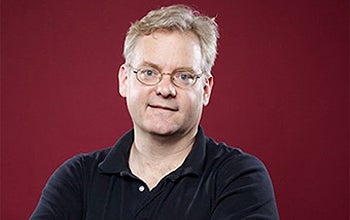
Richard Roberts, professor of chemistry, chemical engineering and materials science, and biological sciences. Photo by Max S. Gerber.
On the other hand, cancer treatment usually involves a combination of drugs to attack the disease from multiple angles. In order to administer multiple drugs at the same time, each drug needs to be relatively nontoxic on its own so that the combination does not injure or kill the patient.
“One of oncology’s main preoccupations is to make drugs that are as selective as possible,” Roberts said. “If we could link our compounds to drugs that are highly toxic to cancer cells, and attack them exclusively, it would probably not affect nearby tissue.”
Successful tests
Roberts’ team has successfully imaged tumors in mice as part of preclinical trials. This is done by taking cells from a human tumor, injecting them into a mouse and waiting for the tumor to grow.
“The way we practice medicine today, there is a lot of emphasis on disease prevention and early detection,” said Uttam Sinha, vice chair of otolaryngology at the Keck School of Medicine of USC, who is working on this project with Roberts. “Our nanoscale diagnostics that use nanopeptides are designed to achieve this goal.”
Although one of the most important parts of the research is actually making the tumor fluoresce, another significant challenge is to avoid inadvertently targeting the lungs, liver, heart and other vital organs.
“It’s not only about hitting the target, but about not hitting everything else,” Roberts said.
However, Roberts’ vision goes far beyond imaging. In the future, he would like to explore ways in which his compounds can be used to actually attack cancer cells as well as small-scale radiation that can target the nuclei of previously identified cancer cells without affecting their surroundings.
“The next step would be trying to kill the tumors,” Roberts said.