The Waiting Is Over
Synapses, the links between neurons that allow them to pass information to one another, are central to all the brain’s functions. These key building blocks are believed to be where memories are stored, yet researchers have little understanding of how they change when memories are made. New insights into the role of synapses in memory, learning and forgetting — or how sleep, drugs and other stimuli affect them — would revolutionize scientific understanding of the entire brain.
At present, researchers cannot image the entire brain at a high enough resolution to see synapses, tiny structures only a hundredth the diameter of a human hair. That’s partly because scientists have yet to build the technology needed to selectively label, image and analyze these key but miniscule structures. MRI, EEG’s and even implanted electrodes can offer only crude data on the brain’s workings.
USC Dornsife’s Don Arnold and Scott Fraser as well as Carl Kesselman of the USC Viterbi School of Engineering, aim to change that.
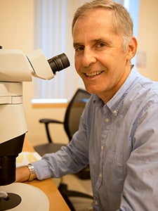
Don Arnold, professor of biological sciences. Photo by Peter Zhaoyu Zhou.
The trio has received a highly competitive five-year, $9.7-million Transformative Research Grant from the National Institutes of Health to perform the first direct study of living synapses in the intact brain, allowing researchers to analyze synaptic changes in zebrafish as a memory is made.
The USC researchers chose zebrafish because their transparent brains make them easier to image. They are also large enough to show the same complexities, mechanisms and diseases as the human brain, including mental illness, making the results relevant to neuroscientists and other health researchers.
“We want to create a new way of observing how the brain functions in a way that nobody has ever done before,” Kesselman said. “Our approach will offer an overall map of the brain’s synaptic changes, creating an entire circuitry diagram compared to just a view of a single wire.”
Arnold, Fraser and Kesselman believe that such a comprehensive picture of the brain’s synapses could one day lead to a better understanding of and treatments for post-traumatic stress disorder (PTSD) and other neurological conditions in humans.
The zebrafish experiments will use a new class of genetically embedded probes developed by Arnold, professor of biological sciences at USC Dornsife, and his team. They have developed a powerful new approach: the zebrafish itself makes a protein that has been engineered to “label” its own synapses with a fluorescent protein that can be seen with the right microscope.
Fraser, with appointments in USC Dornsife, USC Viterbi and the Keck School of Medicine of USC, is working with an interdisciplinary team to build a powerful light sheet microscope optimized to image the synapses that Arnold’s team will label. The microscope is so fast and sensitive that it can capture a comprehensive image of a zebrafish’s synapses before, during and after a memory is made. This would allow researchers to watch the brain in action as the animal responds to various stimuli.
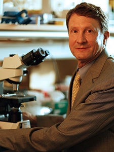
Provost Professor of Biological Sciences and Biomedical Engineering Scott Fraser. Photo by John Livzey.
“We pride ourselves on building microscopes that redefine the impossible,” said Fraser, one of the world’s foremost imaging experts. “This new microscope opens a new doorway in microscopy, perhaps the only tool that is adequate to fully exploit the advantages of the labels Professor Arnold has developed.”
Kesselman — a computer scientist with joint appointments at USC Viterbi and the Keck School — has designed algorithms that will identify, sort and store zebrafish synapses, a challenging proposition given their small size, large number and the size of the data. These huge databases of information “will be accessible to researchers using the ‘big data’ tools the USC researchers are developing,” he said.
To measure synaptic changes when a zebrafish makes a memory, Arnold and Fraser will exploit some of the classical tools used by psychologists to measure learning and memory. For example, they will pair a flashing light with an electric shock until the fish associates the light with discomfort.
By imaging the fish’s synapses before, during and after the learning experience, the team will create a map of brain changes with learning. Similarly, the USC trio will chart synaptic changes caused from the loss of a memory — flashing the light several times but no longer following it with a shock — so that the fish no longer remembers the connection between light and discomfort.
Eventually, researchers will have several highly detailed pictures of synaptic changes resulting from different stimuli, which, in turn, should provide insights into fundamental processes underlying learning and memory.
“People have wanted to see this [synaptic changes] for the last 100 years, but the technology wasn’t there,” Arnold said. “Until now.”